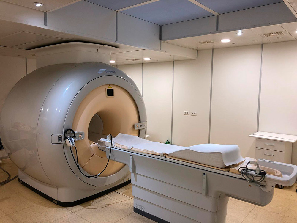
Non-invasive and non-radioactive nature, MRI/MRS provides an ideal approach for diabetes research in humans and animal models.
By virtue of its non-invasive and non-radioactive nature, in vivo magnetic resonance imaging and spectroscopy (MRI/MRS) provides an ideal approach for diabetes research in humans and animal models. Particularly, recent technical advances of in vivo MRI and MRS have made it possible to further understand human metabolism, such as in vivo glycogen metabolism and ATP flux in real time, which may be quite difficult to be assessed by other methodologies. Currently, MRI and MRS methods have been recognized as one of the most powerful tools in diabetes and metabolism research in vivo.
Aims
- Utilization of in vivo MRS and MRI in order to further unravel the pathogenesis and etiology of diabetes and metabolism diseases, such as mechanisms of insulin resistance
- Development of new MR techniques to comprehensively understand questions related to diabetes and metabolic diseases
In essence, we assess content of visceral, muscular and liver fat, ATP and glycogen in liver and muscle, and ATP flux in the tissue in vivo, in conjunction with specific physiologic questions. The state-of-the-art 3T human Philips scanner at DDZ is fully dedicated for this type of research. Primarily, we will focus on 1H-MRS and multinuclear spectroscopy [31P- and 13C-MRS], and also employ additional effective imaging modalities if needed.
Team
Selected Publications
- Jonuscheit M, Wierichs S, Rothe M, Korzekwa B, Mevenkamp J, Bobrov P, Kupriyanova Y, Roden M, Schrauwen-Hinderling VB 2024. Reproducibility of absolute quantification of adenosine triphosphate and inorganic phosphate in the liver with localized 31 P-magnetic resonance spectroscopy at 3-T using different coils. NMR Biomed. 37. https://doi.org/10.1002/nbm.5120
- Jonuscheit M, Uhlemeyer C, Korzekwa B, Schouwink M, Öner-Sieben S, Ensenauer R, Roden M, Belgardt B-F, Schrauwen-Hinderling VB 2024. Post mortem analysis of hepatic volume and lipid content by magnetic resonance imaging and spectroscopy in fixed murine neonates. NMR Biomed. 37. https://doi.org/10.1002/nbm.5140
- Michelotti FC, Kupriyanova Y, Mori T, Küstner T, Heilmann G, Bombrich M, Möser C, Schön M, Kuss O, Roden M, Schrauwen-Hinderling VB 2024. An Empirical Approach to Derive Water T1 from Multiparametric MR Images Using an Automated Pipeline and Comparison With Liver Stiffness. J Magn Reson Imaging. 59: 1193-1203. https://doi.org/10.1002/jmri.28906
- Kupriyanova Y, Zaharia O-P, Bobrov P, Karusheva Y, Burkart V, Szendroedi J, Hwang JH, Roden M for the GDS Group 2021. Early changes in hepatic energy metabolism and lipid content in recent-onset type 1 and 2 diabetes mellitus. J Hepatol. 74: 1028-1037. https://doi.org/10.1016/j.jhep.2020.11.030
